
#Celiac disease colonoscopy findings free#
Seen in patients on gluten free diet (suggesting minimal amounts of gluten or gliadin are being ingested) patients with dermatitis herpetiformis family members of celiac disease patients, not specific, may be seen in infections.

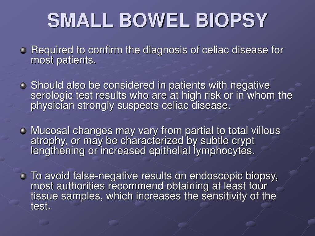
Grade A/Type 1: increased intraepithelial lymphocytes but no villous atrophy.Simplified systems may be more reproducible (Corazza, Roberts, Ensari) Type 3: Spectrum of changes seen in symptomatic celiac disease.Type 2: Very rare, seen occasionally in dermatitis herpetiformis.pylori, hypersensitvity to food, infectons (viral, parasitc, bacterial), bacterial overgrowth, pharmacological drugs (mainly NSAIDs), IgA defcit, common variable immunodefciency or Crohn’s disease. This may also indicate gastroduodenits caused by H. Type 1: Intestinal lining has been infiltrated with IELS – seen in patients on a gluten free diet (suggesting minimal amounts of gluten or gliadin are being ingested), patients with dermatitis herpetiformis and family members of celiac disease patients.Type 0: Intestinal lining is normal -celiac disease highly unlikely.*IEL/100 enterocytes – intra-epithelial lymphocytes (IELS) per 100 enterocytes (epithelial cells in the small intestine) The endoscopy itself may show scalloping and/or flattening of dudodenal folds, fissuring over the folds, and a mosaic pattern of mucosa of folds. Epithelial changes, including structural abnormalities in epithelial cells.The lamina propria is a thin layer of loose connective tissue, which together with the epithelium forms the mucosa which stops pathogens from entering the body. Mononuclear cell infiltration in the lamina propria.This is a shrinking and flattening of the villi due to repeated gluten exposure. Crypt hyperplasia is when the grooves are elongated compared to a normal intestinal lining which has short crypts. Crypts are grooves between the villi, which are the small fingerlike projections that line the small intestine and promote nutrient absorption. Crypt hyperplasia with a decreased villi/crypt ration.Epithelial cells line your intestines and act as a barrier between the inside and the outside of your body. More than 25 IELs per 100 epithelial cells is significant. Density of intra-epithelial lymphocytes (IELs), which are white blood cells found in the immune system.What Does the Intestinal Biopsy Show? (Give Me the Science) It is recommended that the doctor take at least 4-6 duodenal samples from the second part of duodenum and the duodenal bulb, in order to obtain an accurate diagnosis. Samples of the lining of the small intestine will be studied under a microscope to look for damage and inflammation due to celiac disease. A scope is inserted through the mouth and down the esophagus, stomach and small intestine, giving the physician a clear view and the option of taking a sample of the tissue. An endoscopy is a procedure that allows your physician to see what is going on inside your GI tract. If the results of the antibody or genetic screening tests are positive, your doctor may suggest an endoscopic biopsy of your small intestine. An intestinal (duodenal) biopsy is considered the “gold standard” for diagnosis because it will tell you (1) if you have celiac disease, (2) if your symptoms improve on a gluten-free diet due to a placebo effect (you feel better because you think you should) or (3) if you have a different gastrointestinal disorder or sensitivity which responds to change in your diet.


 0 kommentar(er)
